Chest X-Ray
A chest X-ray is a test that produces black and white images of the body parts inside your chest.
What is a chest X-ray?
Why do I need a chest X-ray?
What are the risks of chest X-rays?
How do I prepare for a chest X-ray?
What happens during a chest X-ray?
What happens after a chest X-ray?
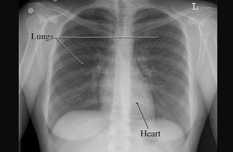
What is a chest X-ray?
A Chest X-ray is a test that produces black and white images of your body parts, including:
- Heart
- Lungs
- Blood vessels
- Spine and chest bones
- Air and fluid in or around your lungs
Why do I need a chest X-ray?
A Chest X-ray can identify the cause of symptoms like:
- Shortness of breath
- Ongoing cough
- Chest pain
- Chest injury
A Chest X-ray can also help your doctor to work out if you have:
- Chest infections
- Heart failure
- Fluid or air collection around the lungs
Chest X-rays help to check the position of wires, leads and tubes after heart procedures and surgery.
What are the risks of chest X-rays?
Chest X-rays are painless procedures, and they have very few risks. You may be concerned about chest X-rays as there is radiation involved. However, the amount of radiation used is very low and isn’t harmful.
How do I prepare for a chest X-ray?
You won’t need to do any special preparation for your chest X-ray in the days leading up to the test. Just before the procedure, you’ll be asked to wear a hospital gown and remove all jewellery - including body piercings and glasses.
What happens during a chest X-ray?
During a chest X-ray, you’ll be asked to stand in front of an X-ray machine and press your body against an X-ray plate. The X-ray technician may ask you to stand still and hold your breath while the images are taken - this helps to provide clearer pictures. Pictures will be taken from the back and from the side, so you may be asked to move into different positions. The procedure takes less than 10 minutes.
What happens after a chest X-ray?
When your chest X-ray is finished, a specialist (radiologist) will study the images and prepare a report for your doctor. Your doctor will then discuss your results and recommend the best treatment plan for you.